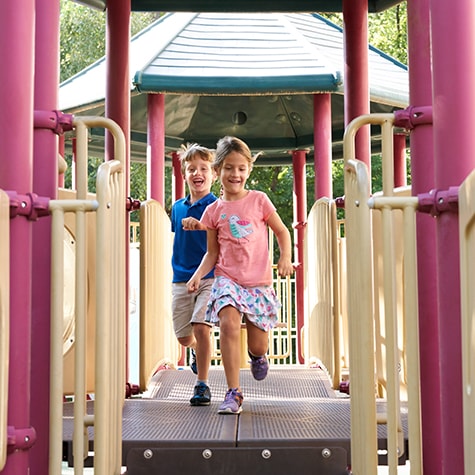
Bones—no matter how big or small—help protect a child’s internal organs and her muscles. Strong, healthy bones help children participate in their favorite activities, such as dancing, baseball, recess and simply playing in the backyard with friends.
Some children develop bone diseases as they grow. These diseases can be genetic or be caused by a lack of vitamins or minerals in the bone. Taking your child to a pediatric orthopedist is key to treatment that caters to your kid’s needs in the event of a condition that may cause damage to your child’s bones.
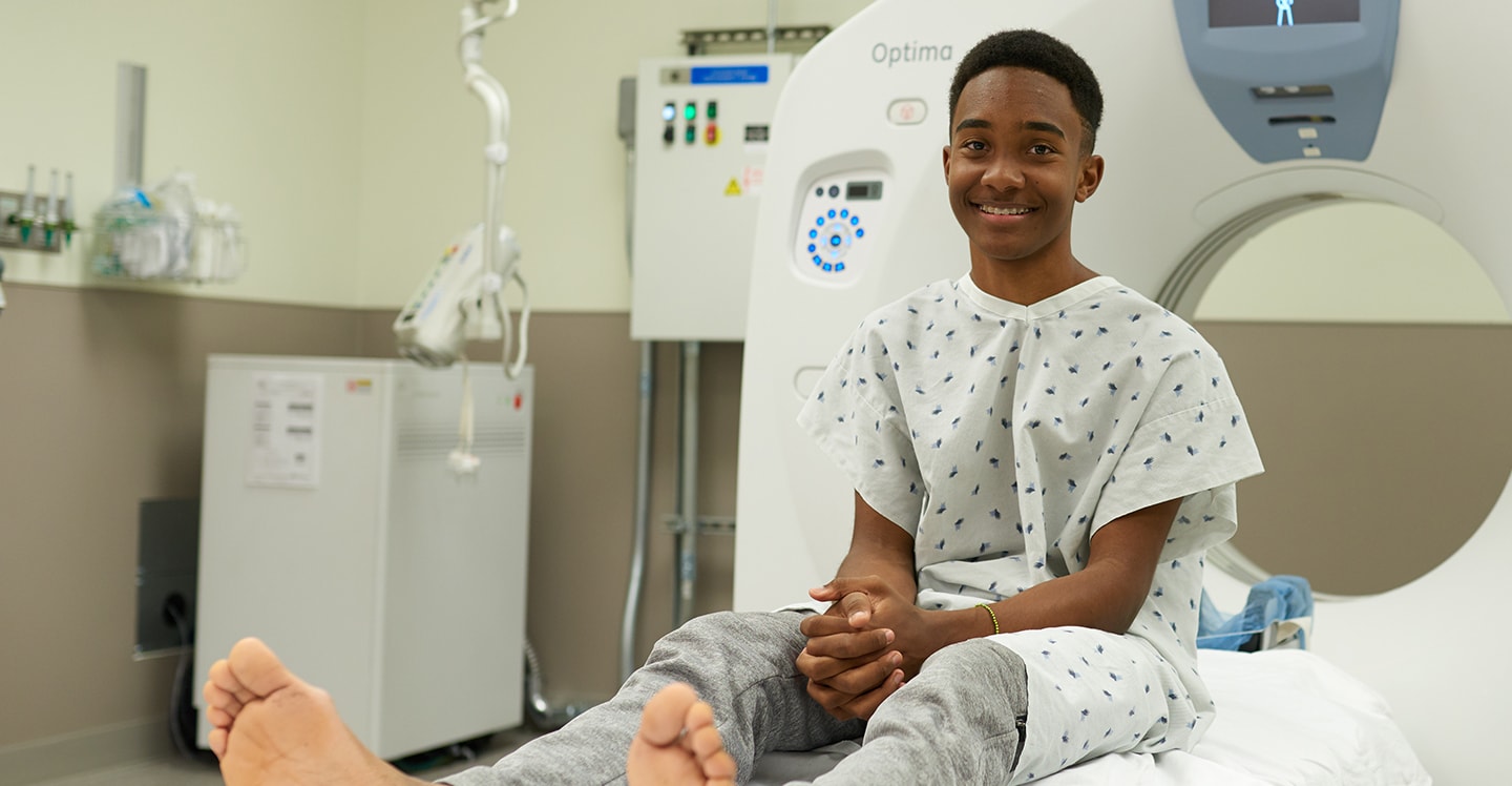
Our pediatric bone specialists know how to treat bone disease in growing kids and teens.
Our pediatric bone specialists know how to treat bone disease in growing kids and teens. We also work closely with our endocrinology bone health specialists to offer your child comprehensive care. When it comes to bone health, it’s important to remember that children are not just small adults. Kid bones differ from adult bones in significant ways and that’s why they need our care.
Bone is made of a soft, organic portion (collagen) and hard, inorganic portion (minerals). Collagen is a strong protein material in which fibers are woven together to provide bone its tensile strength. Minerals principally made of calcium and phosphate, which provides a bone its strength.
Bones store 99% of the body’s calcium. If the body does not have enough calcium from the diet, then it will take calcium from the bone to continue functioning normally.
Vitamin D is a hormone that enables the intestines to absorb calcium from the diet into the bloodstream. Like skin, bone is continually refreshing its cells. During childhood, bone formation occurs at a rate faster than bone loss. Childhood is the only time when bones gain mineral and become stronger. This is why it is crucial to maximize your child’s ability to gain bone mineral and decrease the chance of developing osteoporosis as an adult. Between 20 to 60 years old, bone is essentially at a steady state in which bone formation and bone breakdown are occurring at equal rates. As we get older, bone loss occurs faster than the body can generate new bone.
Approximately 60-80% of our bone mineral density is determined by our genes. Because of this, we can control only a small portion of our bone mineral makeup. Diet, exercise, chronic illness and smoking can impact long-term bone health. Foods rich in vitamin D and calcium include plain yogurt, calcium-fortified orange juice, mozzarella, fish (including sardines, canned tuna and canned salmon), milk, tofu made with calcium sulfate and cottage cheese. Children’s is focused on optimizing our young patients’ bone health.
There are many conditions that can affect a child’s bone health. And since a child’s or teen’s bones are still growing, and conditions and treatments can impact kids’ bones differently than adult bones, where you take you them matters. Our doctors at Children’s Healthcare of Atlanta understand how to treat many bone conditions in growing kids and teens, including:
Bone Conditions
What is a bone or joint infection?
Bone and joint infections (osteomyelitis and septic arthritis) occur much more commonly in children than in adults, with the majority of cases occurring in children under the age of 5.
What causes bone or joint infections?Bone infections are caused by bacteria in the bone, which is very common in childhood. This can result from other infections, such as from the ear, nose and throat, or it can be from simply brushing the teeth. Most of the time, the immune system wins, and the bacteria is cleared from the blood. But if there is a large amount of bacteria in the blood and it is strong enough, it can infect the bone.
The causes of joint infections are not well understood. Most of the time, the immune system and joint can eliminate a bacterial infection before it overwhelms the body; but if the bacteria is particularly strong and/or large in number, then it may overwhelm the joint and cause an infection. In addition, an infection from a bone can cross into a joint. This commonly occurs in four locations—hip, shoulder, elbow and ankle.
Children will usually have a fever and have a limp on the extremity affected, refuse to move the limb or refuse to walk at all. Most children also have swelling and tenderness at the site of the infection, and the area may be warm.
If the child’s joint is infected, movement may be extremely painful. A combination of blood tests may be helpful in diagnosing the infection. An MRI may also be necessary to identify the exact location of the infection.
How do doctors treat bone and joint infections in kids?Like other infections, antibiotics are a primary treatment. Antibiotics are initially given through an I.V. Once the infection shows significant improvement, your child's doctor may switch to antibiotics taken by mouth. If the infection involves a child’s joint, then surgery may be necessary to “clean out” the joint to prevent cartilage damage. If the infection involves the bone, surgery may be necessary as well.
It is extremely important in the case of joint infections to begin physical therapy immediately to help your child move her joint, as it can become permanently stiff very quickly.
If infections are treated promptly, there are often no long-term consequences. However, infections can weaken the bone, at least temporarily, and this puts your child at risk for a fracture until the bone becomes strong again. For joint infections, cartilage damage can be permanent and lead to pain and future arthritis. It is very important for your child to continue seeing the doctor to help identify potential problems early.
Osteogenesis imperfecta is a genetic disorder that impacts the body’s ability to produce collagen, a protein that helps strengthen bones. It can be a mild bone disorder, resulting in a few fractures in a child’s lifetime. In more severe cases, the disease can cause hundreds of fractures and even impact other organ systems.
There is no known cure for osteogenesis imperfecta. Current treatments focus on minimizing pain, fractures and bony deformities. Our goal is to help children with this condition live better, more fulfilling lives. Treatments range from physical therapy to bracing, medications and occasionally surgery.
What is rickets?
Rickets is a childhood condition in which the bones lack certain minerals. Although they all look the same on X-rays, there are several different types of rickets. The type depends on which mineral is deficient—either calcium, phosphorus or the enzyme that helps create the mineral portion of bone, alkaline phosphatase.
The two most common causes of rickets include:
- Calcium deficiency: The most common cause of calcium deficiency in bone is a lack of vitamin D, not a lack of calcium in the diet. This is because vitamin D allows the body to absorb calcium. If the body does not have enough calcium in the blood, then it will take the calcium out of bone instead. If this cycle continues, bones can become weaker. This can lead to a child developing physical signs of rickets, such as bowed legs or other bone deformities.
- Phosphorus deficiency: Phosphates support the growth and repair of bones and teeth. The most common reason for a phosphorus deficiency in bone is an inherited disorder known as X-linked hypophosphatemic rickets. This disorder is caused by a defect in the PHEX gene, which regulates the movement of phosphate compounds from the kidney. Normally, the kidneys send phosphates to the bone through the blood. The PHEX gene defect causes the kidneys to get rid of the phosphate instead of sending it through the blood, leading to a phosphorus shortage in the blood. Similar to low calcium in the blood, when the body recognizes there is not enough phosphorus in the blood, it will remove it from the bone instead. This genetic defect may also prevent the kidneys from processing vitamin D, which is necessary for the body to absorb calcium. Girls are twice as likely as boys to have the X-linked hypophosphatemic disorder.
When there is less calcium or phosphorus in bone, the bone and bone’s growth plate become weak. This weakening of bone can lead to slow growth. In severe cases, children can have seizures from calcium deficiencies or develop fractures in weakened bone.
How do doctors treat rickets in kids?
Treatment for rickets depends on which mineral deficiency your child has. This may require the assistance of an endocrinologist. Once your child’s doctor determines the mineral deficiency, treatment involves replacing the mineral—whether it is calcium, vitamin D, phosphorus or a combination of these. In many cases, the bone changes that happen with rickets get better over time. In more severe cases, children may continue to have slowed bone growth and bones are somewhat crooked. Occasionally, bones become so crooked they require corrective surgery. Surgery is followed by bracing for correction.
What is skeletal dysplasia?
Skeletal dysplasia is a group of rare genetic disorders that affect a child’s bones and joints and can hinder her growth and development. Conditions associated with skeletal dysplasia include spinal stenosis, bowed legs, stiff joints or clubfoot.
Signs of skeletal dysplasia may include abnormal growth in your child’s spine and skull, as well as:
- Vision problems
- Cleft palate
- Hearing loss
- Brittle teeth and bones
- Hydrocephalus
- Cervical medullary compression
- Large head with a prominent forehead
- Short stature
- Long trunk with shorter arms and legs
How do doctors diagnose skeletal dysplasia in kids?
Orthopedic specialists will often diagnose the disorder using an X-ray, CT scan, ultrasound or MRI. Your child may also need an eye exam.
How do doctors treat skeletal dysplasia in kids?
Treatment will depend on the specific skeletal dysplasia condition your child is diagnosed with but can include surgeries such as:
- Limb lengthening
- Guided growth
- Surgery to reposition the bone
- Realigning the joint
Conditions That Affect Bone Formation and Growth
Achondroplasia is a shortness in height as a result of a genetic or medical condition.
Infantile Blount’s disease is the most common reason for a bowlegged deformity. In this disease, the growth plate stops growing on the upper, inner portion of the tibia bone. This results in a bowleg deformity that gets progressively worse. Blount’s disease is usually diagnosed around age 2. Infantile Blount’s disease is thought to be a developmental problem rather than a congenital one. Adolescent Blount’s disease, also known as juvenile Blount’s disease, is very different from infantile Blount’s disease. While infantile Blount’s is diagnosed soon after walking, adolescent Blount’s is usually diagnosed when the child is age 10 or older. Children who are diagnosed with adolescent Blount’s have normal-appearing limbs when they are younger, but they develop bowlegs as they become older. While the disease often affects both sides, one side may be more severely bowlegged than the other. In adolescent Blount’s disease, the initial bowlegged deformity tends to be less severe than infantile Blount’s disease, but it has the potential to become just as severe if left untreated. Although some patients with adolescent Blount’s disease may be normal weight, many children with this condition are overweight or obese.
Bowlegs is a condition in which the knees point away from the center of the body. Many young children normally have bowlegs that correct at about 2 years old. This is referred to as physiologic bowlegs. However, there are two conditions in which bowlegs do not correct in young children: infantile Blount’s disease and rickets. Both of these problems can be diagnosed with an X-ray. If your child still has bowlegs by age 2, it is important to have him evaluated for underlying conditions that may be causing the bowlegs.
In this condition, the angle between the top of the femur (thigh bone) and the hip joint is too small. This results in the top of the femur pointing down toward the body. Coxa vara is a decrease in the angle of the top part of the femur. It involves the neck of the femur, which is the part between the ball portion at the top of the femur (head) and the long, straight portion of the femur (shaft). Typically, the angle between the neck of the femur and the shaft of the femur is 135 degrees. When a child has coxa vara, this angle is fewer than 120 degrees.
This is a condition in which a baby is born with her knee and femur (thigh bone) not connected. The leg is stuck in a straight position.
A fracture is a break in the bone that can affect bone growth and health. Learn more about fractures and how we treat them at Children's.
Intoeing means that a child’s feet turn inward when walking or running, instead of pointing straight. It’s commonly referred to as being pigeon toed.
Knock knees is a condition in which the knees point inward and touch. In most cases, knock knees resolve by about 6 years old. However, if the crookedness does not resolve on its own and continues to worsen, treatment may be needed to correct the deformity.
Bone Cysts and Tumors
What is a simple or unicameral bone cyst?
A unicameral bone cyst, also known as a simple bone cyst, is a benign (noncancerous) bone tumor. These tumors are mostly commonly found near the shoulder and hip joints. They often go away without treatment after your child has completed growing.
Treatment can range from observation to surgically removing the cyst. Although simple cysts are benign and usually go away with time, they can still lead to problems. They have the potential to weaken bone and can lead to breaks, which can be painful and may require surgery.
How do doctors diagnose bone cysts in kids?
These cysts are usually found in one of three ways:
- Bone fracture: Sometimes these fractures can be treated with a cast, and the cyst can be observed later. Other times, the fracture can involve critical areas like the hip. In these cases, your child's surgeon may recommend surgery to prevent future growth complications.
- Complaint of pain: If your child has pain in an area, an X-ray may reveal a cyst. If a cyst can be seen on an X-ray, it is likely that a fracture is about to occur. This may be the cause of your child’s pain. Surgery to remove the cyst may be required to prevent the fracture.
- Incidentally: Occasionally, X-rays, an MRI, CT scan or biopsy may be performed for one reason, and an otherwise asymptomatic cyst—or a cyst with no prior symptoms—will be detected. Treatment options can vary based on the child and doctor.
How do doctors treat bone cysts in kids?
Cysts have often been treated by injecting them with steroids to reduce the size. However, when using this method, the vast majority (approximately 70%) of cysts return. We limit the risk of a cyst returning by “scraping out” and then filling the space with injectable calcium using minimally invasive surgery. The calcium is used to help stimulate healing. This method decreases the recurrence rate of cysts by about 30%. Once your child has been treated for a cyst, it is important she continue to see the doctor, even if there is no pain, to help ensure the cyst does not return.
What is an osteochondroma?
The most common noncancerous bone growth, osteochondroma is an overgrowth of cartilage and bone that happens at the end of the bone near a child’s growth plate. It typically affects the long bones in the legs, pelvis or shoulder blades and impacts children age 10 through adults age 30.
What causes an osteochondroma?
The exact cause of osteochondroma is unknown, but symptoms may include:
- Shorter than normal height for a child’s age (if associated with other conditions).
- One leg or arm that is longer than the other (if associated with other conditions).
- Pressure or irritation when your child exercises.
- Soreness of the muscle near a bone.
- A hard mass that is painless and doesn’t move.
How do doctors diagnose an osteochondroma?
Osteochondroma is usually diagnosed with an X-ray, CT scan or MRI and is treated based on a child’s age; overall health and medical history; how sick a child is; how well a child can handle specific medications, therapies or procedures; and how long the condition is expected to last. Treatment may include medication or surgery.
How do doctors treat osteochondroma?
Treatment of osteochondroma varies depending on the symptoms, age of the patient and any other associated deformities.
What are sarcomas?
Sarcomas are bone and soft tissue cancers that impact connective tissue (tendons) and muscles. They are a rare condition in children, accounting for fewer than 3,000 new cases annually in the U.S. and approximately 11% of all tumors. The two most common sarcomas in a growing child are osteosarcoma and Ewing’s sarcomas.
Advances in imaging and chemotherapy have dramatically increased the long-term survival of patients diagnosed with sarcomas. MRI has also improved our ability to see the tumor and evaluate its response to chemotherapy.
How do doctors diagnose sarcomas or tumors in kids?
The doctor will X-ray and MRI the entire area, including the joint above and below the tumor. The doctor may then recommend a CT scan of the chest to view if there is cancer (metastatic lesions) or perform a bone scan to identify other conditions (skip lesions or other bony metastases). Your child may also need a bone marrow biopsy to determine if there is soft tissue or Ewing’s sarcoma.
How do doctors treat sarcomas in kids?
Chemotherapy and radiation have resulted in an increased survival rate. With pre-operative chemotherapy and surgery, the five-year survival rate for osteosarcoma and Ewing’s sarcoma approaches 70-80%. Genetics advancements are still evolving and will likely play a significant role in treatment in the near future.
Surgical treatments, also known as local control, involve removing the cancer. Surgical treatment options depend on the size, location and involvement of surrounding structures (whether the cancer has spread to other areas). Surgery options may include:
- Simple resection
- Limb-sparing or limb-salvage
- Amputation
- Rotationplasty
Children who require surgery to remove the tumor and chemotherapy or radiation to treat the cancer, may require physical therapy to restore range of motion, strength, and overall conditioning and function. Some children will require prosthetics following an amputation or a brace (orthotics) to protect the limb.
When there’s an underlying issue with your child’s bones, Children's physicians use the most advanced techniques to diagnose and treat your child.
We provide:
- Unique and specialized expertise in treating bone disease.
- Cutting-edge research in diagnosing musculoskeletal infections (joints, ligaments, muscles, nerves, tendons, and structures that support the limbs, neck and back).
- Nonsurgical treatments, such as antibiotics and supplements.
- Minimally invasive techniques to treat most bone cysts.
- Nationally recognized treatment for children with sarcomas (bone cancers).
- Innovative techniques and the newest implant equipment for limb-salvage surgery.
Our New Osteogenesis Imperfecta Clinic
Our multidisciplinary Osteogenesis Imperfecta (OI) Clinic brings together specialists from orthopedics, otolaryngology (ENT), pulmonology, endocrinology, dentistry, orthotics, and physical and occupational therapy to provide the most comprehensive care possible for patients with OI. Our clinic staff is specially trained to care for growing kids and teens with OI, from birth to age 21.
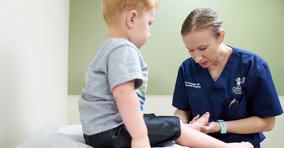
Make an AppointmentOur pediatric-trained orthopedic surgeons use advanced techniques to diagnose and treat children with bone disease.

Building a campus to support healthcare for generations to come
Scheduled to open fall 2024, the North Druid Hills campus’ new hospital will be dedicated to advancing orthopedic care for children of all ages—birth to 18.
LEARN MORE-
Athens Orthopaedics
1181 Langford Drive, Building 200, Suite 101
Watkinsville, GA 30677 -
Buford Orthopaedics
2914 Vinson Court
Buford, GA 30518 -
Center for Advanced Pediatrics
1400 Tullie Road NE
Atlanta, GA 30329 -
Children’s at Cherokee
1558 Riverstone Parkway, Suite 100
Canton, GA 30114 -
Children’s at Duluth
2270 Duluth Highway 120
Duluth, GA 30097 -
Children’s at Fayette
1265 Highway 54 West, Suite 200
Fayetteville, GA 30214 -
Children’s at Forsyth
The Collection at Forsyth
410 Peachtree Parkway
Cumming, GA 30041 -
Children’s at Hudson Bridge–Orthopaedics and Sports Medicine
1492 Hudson Bridge Road
Stockbridge, GA 30281 -
Children’s at Meridian Mark
5445 Meridian Mark Road NE
Atlanta, GA 30342 -
Children’s at Old Milton Parkway
3300 Old Milton Parkway
Alpharetta, GA 30005 -
Children’s at Town Center Outpatient Care Center
605 Big Shanty Road NW
Kennesaw, GA 30144 -
Children’s Orthopedics and Sports Medicine – Douglasville
Opening September 6, 2022
6095 Professional Parkway,
First Floor, Suite 101B
Douglasville, GA 30134 -
Macon
4660 Riverside Park Blvd.
Macon, GA 31210
Contact Us 404-255-1933