At Children’s Healthcare of Atlanta, we use advanced diagnostics, treatment and neuroimaging technology to provide better outcomes for your child. Our team provides unique treatment options based on each child’s condition and needs. Below is a list of evaluations and diagnostic tests your child may undergo or be prescribed during his treatment journey. These explanations aim to give your family a general understanding of the tests and/or technology employed. Talk to your child’s clinical care team for more information.
- Ambulatory electroencephalogram (EEG): An ambulatory EEG measures the electrical signals of your child’s brain while he’s doing his everyday activities, such as playing with friends, watching TV and sleeping. Your child will have sensors on his scalp and carry a backpack with special equipment that records his brain signals. Unlike a traditional EEG, an ambulatory EEG is done outside a clinic or hospital.
- Angiography: An angiography is an imaging test that uses X-rays to view blood vessels in the body, specifically narrow, blocked, enlarged or malformed arteries, or veins in your child’s brain, heart, abdomen and legs. The images are called angiograms.
- Dense array EEG: A dense array EEG measures and records the brain’s electrical activity by using up to 256 electrodes. By placing this many electrodes on key points on the patient’s head, doctors are able to get a good approximation of where your child’s seizures start.
- Diffusion tensor imaging (DTI) fiber tracking: DTI uses computer technology to see the complex network of nerve fibers connecting the different areas of a child’s brain. This technology can help surgeons plan for surgery.
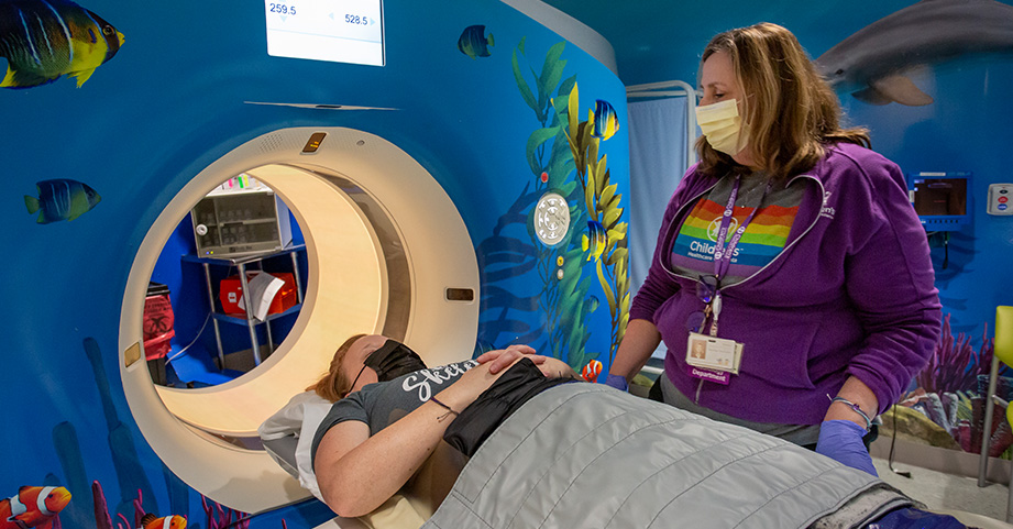
- CT scan: The CT scanner is a doughnut-shaped machine used to view parts of a child’s body that cannot be seen with a regular X-ray.
- Cortical stimulation (also called corticography or cortical mapping): Cortical stimulation is often used before surgery to help a neurosurgeon make decisions about what areas of the brain are affected by seizures. This process involves the placement of a grid on your child’s brain. This grid has electrodes to help measure the electrical activity on the surface of the brain.
- EEG: An EEG is a test that measures the electrical activity in the brain, called brain waves. A routine EEG happens at the hospital or in a doctor’s office.
- Electromyography (EMG): An EMG is a test that is used to record the electrical activity of muscles.
- Fluorodeoxyglucose (FDG)-PET/MRI co-registration: FDG-PET/MRI co-registration is used to help plan a child’s surgery. We are one of only a few centers in the country that uses FDG-PET/MRI co-registration for pediatric epilepsy surgery.
- Functional MRI (fMRI): An fMRI is a technology that takes advantage of measuring functional skills through the use of MRI technology.
- Intra-operative MRI (iMRI): The iMRI produces high-quality images that allow surgeons to better identify tumors and lesions during surgery.
- Lumbar puncture (spinal tap): A lumbar puncture may be used to get a sample of the fluid that surrounds your child’s brain and spine in order to test for serious infections or disorders. This may also be used to give patients medication.
- MRI: An MRI produces very clear images of the human body without the use of X-rays. Our pediatric MRI uses a large magnet, radio waves and computers to take pictures of your child’s body.
- Magnetic resonance spectroscopy (MRS): MRS is a tool used to measure chemical changes in a child’s brain. This tool is combined with an MRI and can compare normal brain tissue to abnormal tissue. It can be used to look at tumors, strokes and epilepsy.
- Mock scanner: A mock scanner allows patients to undergo practice MRI sessions with clinicians and child life specialists, helping to reduce a child’s anxiety before going into the real scanner.
- Morphometry: Morphometry compares the cortical thickness of various brain regions in children with and without neurological disorders. Developmental variation and changes in the cortical thickness can tell us about your child’s development and the impact of a disease or disorder.
- Neurocritical Care Intensive Care Unit (ICU) monitoring: Using a video electroencephalogram (vEEG), our team can monitor neurocritical care patients in our ICU 24 hours a day. We can detect real-time nonconvulsive seizures, which occur in about 30 percent of our monitored patients. Because these seizures are nonconvulsive, they are hard to detect. Our monitoring unit allows us to see these seizures and provide treatment.
- Nerve conduction velocity (NCV): An NCV is a test to see how fast electrical signals move through a nerve.
- Positron emission tomography (PET): PET imaging offers physicians a unique view of the body’s organs and tissue because it records function by showing pictures at the cellular level.
- Single photon emission computed tomography (SPECT): A SPECT is a nuclear imaging scan that integrates CT and a radioactive tracer that allows doctors to see how blood flows to tissues and organs in your child.
- Subtraction ictal single photon emission computed tomography co-registered to MRI (SISCOM): SISCOM is a tool used with an MRI to help find parts of the brain that are having seizures. This tool helps our team plan epilepsy surgeries for pediatric patients.
- vEEG: A vEEG combines an EEG with video of a patient. Our Epilepsy Center has 12 advanced, all-digital vEEG beds, allowing physicians to constantly record and later monitor our patients’ conditions.
Having a child diagnosed with a neurological condition can be an emotional and overwhelming experience. At Children’s, our No. 1 priority is to support you and your family. Whether treating a toddler during an emergency or helping a teen through recovery after surgery, we make it our mission to provide the best care—and best experience—for every child. Family is a big part of your child’s well-being. Not only are you a vital member of your child’s healthcare team; you are a source of security and comfort.

We work to support your whole family while your child is in our care—and after she goes home.
Contact Us 404-785-KIDS (5437)